
Echocardiogram
At Hunterdon Cardiovascular, when we want to get a better idea of how your heart is beating and pumping blood, one of the first tests we perform is an echocardiogram. This test doesn’t involve any radiation and is a basically risk-free way to check for signs of heart disease.
What is an echocardiogram?
The name echocardiogram gets its name because it uses high-frequency sound waves to produce images of your heart’s chambers, valves, walls, and the blood vessels that are attached to your heart (aorta, arteries, veins). Sometimes these tests are called diagnostic cardiac ultrasound or just “echo.”
How does an echocardiogram create these images?
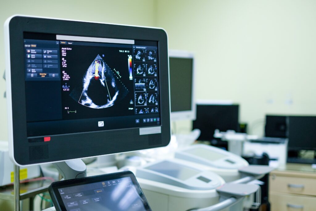
Your Hunterdon sonographer takes the handpiece/probe, called a transducer, and passes it across your chest. The probe sends sound waves down into your chest. These waves bounce off your heart and return to the probe. They “echo” back, if you will. The computer associated with the system then assembles this information into pictures that are viewed in real-time on a video monitor next to the patient.
Who is a candidate to have an echocardiogram and what is measured?
These tests enable your Hunterdon Cardiovascular team to get a look at your heart’s structure, and the quality of its function. You may be having shortness of breath or chest pains. We may suspect heart disease. These are what we can examine with an echocardiogram:
- The size and shape of your heart
- The size and thickness and movement of the walls of your heart
- Your heart’s pumping strength
- How your heart moves when pumping
- Heart valve function
- If blood is leaking backwards through the valves
- If the valves are too narrow
- If there is a tumor or growth around your valves
- If there are problems with the outer lining
- If there are problems with the large blood vessels that enter and exit the heart
- If there are blood clots in the chambers
- If the heart has any abnormal holes
What are the different types of echocardiograms?
Transthoracic echocardiogram
This is the standard type of echocardiogram described above.
Transesophageal echocardiogram
- If we need more detailed images, or if we are unable to get a clear picture of your heart with the standard echo, we may move to this type.
- Your throat is numbed and you’re given a sedative.
- A flexible tube containing a transducer is guided down your throat and into the tube connecting your mouth to your stomach, the esophagus.
- The transducer records the sound wave echoes from your heart.
- A computer converts the echoes into detailed moving images of your heart, which are displayed on the monitor adjacent to the examining table.
Doppler echocardiogram
When we focus on the blood flow in your heart, this is called a Doppler echocardiogram. This measures the change in pitch when the sound waves bounce off blood cells moving through the heart and blood vessels. These changes are known as Doppler signals and they allow us to measure the speed and direction of the blood flow in your heart. Doppler techniques are used in both transthoracic and transesophageal echocardiograms.
Stress echocardiogram
In many cases, problems will only show themselves when the heart is working hard, as during physical activity. This is particularly true when looking at flow through the coronary arteries.
- Ultrasound images of your heart are taken when you are at rest.
- Then you walk on a treadmill or ride a stationary bike for a period of time to elevate your heart rate.
- Another set of ultrasound images is taken immediately after your heart rate has been raised
3D Echocardiograms with Strain
Recent technological improvements with echocardiograms have allowed the creation of three-dimensional images of your heart. These images overcome many of the limitations presented by two-dimensional echocardiograms. Real-time 3D echocardiography allows single- or multi-beat data. Strain rate imaging is a way of measuring deformation of the heart muscle during the echocardiogram, usually using Doppler signals.
What are the risks involved with an echocardiogram?
There are no risks involved with standard transthoracic echocardiograms. Sound waves have no negative effect on the human body. With a transesophageal echocardiogram, your throat may be sore for a few hours after the test, but that’s about it. Stress echocardiograms may cause an irregular heartbeat. Serious complications during stress echocardiograms, such as a heart attack, are very rare. These are very low-risk tests.
When will I learn the results of my echocardiogram?
Your Hunterdon Cardiovascular doctor will review the pictures of your heart within 24-48 hours after your test. The results will be prepared in a report that will describe your heart’s anatomy, heart movements, and any defects or abnormalities observed during your test. You will likely know the results of your test within 3-4 days. These will also be shared with your family doctor.
Schedule A Consultation
If you’re interested in learning more about an Echocardiogram, please contact us at (908)-788-1710